Fetal Cardiology

Fetal cardiology is the diagnosis and treatment of heart problems in babies before birth. This very important branch of pediatric cardiology allows for early diagnosis of heart defects and heart rhythm disorders, so that we can be as prepared as possible for the birth of your baby. If a heart issue is found, families are counselled about the optimal care of their child during the pregnancy and after birth. Sometimes, this means planning for delivery at a children’s hospital outside of Central Oregon, so that a team of pediatric subspecialists is available to assist in the care of the baby right after he/she is born.
We are here to help you every step of the way, whether it means providing follow-up care, helping coordinate medical appointments, or providing assistance through the Children’s Heart Fund.
Who should have a fetal echo?
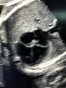
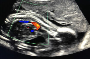 A fetal echo is a special ultrasound that evaluates a baby’s heart anatomy and heart rhythm in detail.
Who should have a fetal echo?
Your obstetrician will decide if you need to have a fetal echo. In general, mothers who are at increased risk for having a baby with a heart abnormality should have a fetal echo. Some of the risk factors are…
- A question of a heart abnormality on a routine ultrasound
- When the baby’s heart rate is too fast, too slow, or irregular
- A family history of a congenital heart defect
- A maternal illness such as diabetes or phenylketonuria
- Certain maternal viral infection such as rubella, toxoplasmosis, CMV, or others
- When the mother has a connective tissue disorder, such as lupus
- Certain medications or drugs taken by the mother, such as anti seizure medicines, lithium, indomethacin, alcohol, amphetamines, cocaine, some antidepressants, and others
- When an obstetric ultrasound detects abnormalities of other organs
- When there is increased likelihood of a genetic syndrome, such as Down syndrome
- When the baby is found to have heart failure on a routine ultrasound, or is not doing well for unclear reasons
- What is the risk of my child having a congenital heart defect?
How accurate is a fetal echo?
In general, fetal echos are very accurate, and can detect nearly all forms of heart disease, including most major heart defects. However, because fetal circulation is different from the baby’s circulation after birth, a few defects may not be found until after the baby is born. Also, some minor defects may not be detected on a fetal echo. The accuracy of the echo is also very dependent on the image quality, movement of the baby, and the baby’s position in the uterus.
Who performs the fetal echo?
What if there is an abnormality?
When should my fetal echo be performed?
 The baby’s heart can be evaluated as early as 16 weeks gestation or anytime after that. It is useful to wait until 20-24 weeks, when the resolution of the echo may be improved.